To truly understand vascular pathology in the brain and in the therapy field you need to have a grasp of the Circle of Willis, the source of blood supply to the brain and the location of over 80% of non-traumatic subarachnoid hemorrhages and the pathway of thromboembolic clots from the heart, internal carotid arteries, and vertebrobasilar system.
Circle of Willis Anatomy
This video by AnatomyZone.com was the first one that I found in my search for better anatomy resources. 2-D anatomy texts just do not compare for those like me who have trouble extrapolating details from text based images. I had to withdrawal from mechanical drafting because I could not draw a 3-D screw from a 2-D image.
Saccular (Berry) Aneurysms
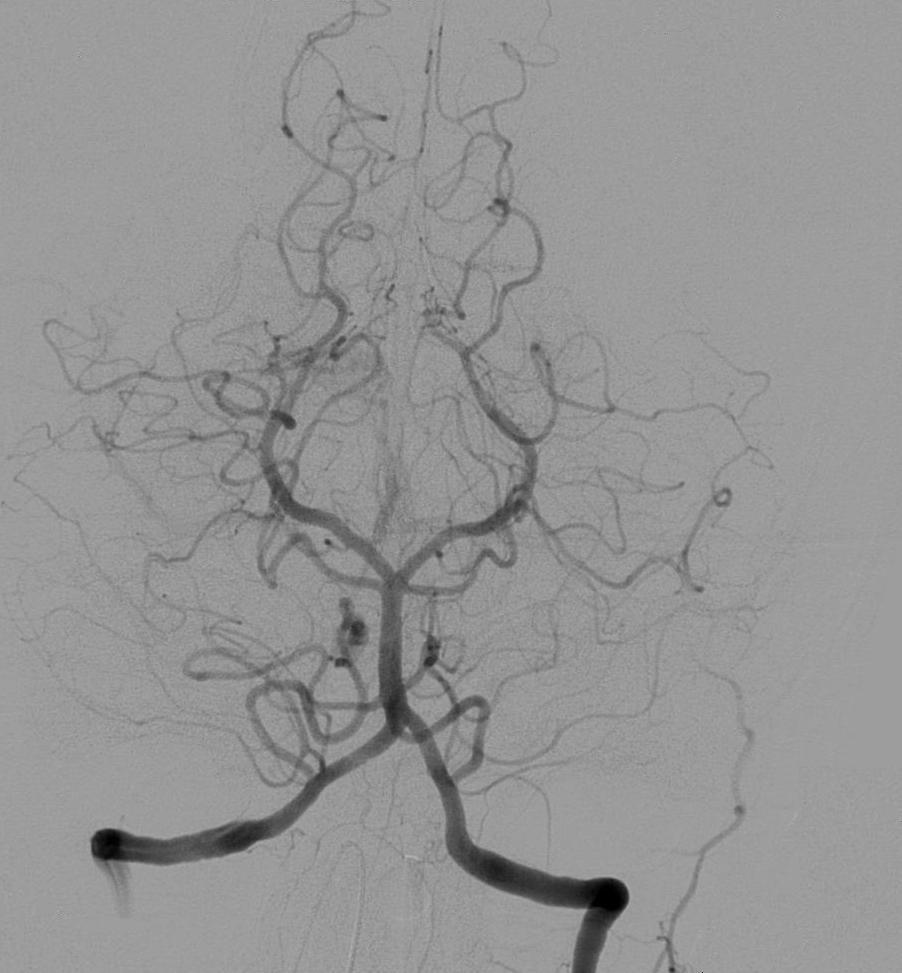
The exact vasculature of the Circle of Willis can be seen by cerebral angiogram.
Berry aneurysms form about 97% of the aneurysms of the central nervous system. They are so named due their appearance as little berries. They are typically formed along the junction or confluence of two anastomosing arteries due to the every constant arterial pressure. These are measured in millimeters a and are at a higher risk of rupture with spikes of hypertension due to pathology, drug use, and exacerbated uncontrolled hypertension. Surgical clipping by a neurosurgeon or coiling with intervential radiology has a distinct roll when the aneurysm has reached 7 millimeters or more.
Saccular (Berry) Aneurysms at the Circle of Willis
Subarachnoid Hemorrhage is a common pathology centered at the Circle of Willis. About 5 percent of strokes are due to SAH. Most people complain of a thunderclap head or the worst headache in their life. As a therapist you will not see individuals at this initial stage, but always keep in mind that it is important to understand the symptoms and causes of your client’s diagnosis. Ignorance almost always causes confusion in a therapist’s treatment plan and recommendations.
Anastomosis of the Circle of Willis
Those who suffer vascular pathology like a thromboembolic clot can have different degrees of insult due to variation in how the vessels anastomose to form the Circle of Willis. Work long enough and you will see clients with a large clot who should not be up walking and talking, but still can due to collateral circulation. To further understand the nature of anastomoses, Anatomy Atlases.org has an illustrated collection of variations of the Circle of Willis.
Take Away
When evaluating your neurosurgical and neurological patients, it is important to realize why they are having functional deficits. Underlying this understanding is your knowledge of neuroanatomy and pathology. While it is true someone suffering from a CVA or aneurysm can present with similar movement deficits, the hospital course, risks, and medical treatments are different. Furthermore, less pronounced functional differences can be spotted when a knowledgeable therapist can predict problems due to type and location of the insult. We are called on to know the difference to be a more complete member of the interdisciplinary team.
additional reference – cited July 3rd, 2014
additional reference – cited July 3rd, 2014
Written By:
Stephen Carnazzo
